Background and Aim:
Neurodegenerative brain disorders like Parkinson’s disease (PD) and Alzheimer’s disease (AD) are affecting more than 50 million people worldwide. Early diagnosis and accurate characterization of the disease progression can be very important for the treatment and the improvement of the patients’ life quality. Currently, PD or AD diagnosis mainly relies on mental status examinations and neuroimaging scans, which are expensive, time-consuming, and sometimes inaccurate.
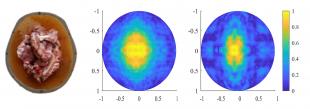
New cost-effective and accurate diagnostic tools and techniques are therefore urgently needed especially for the early detection and prediction of important degenerative disorders (e.g. PD or AD) at the individual level. Over the last decade, microwave medical imaging has emerged as an economical and non-invasive alternative technique for the study of brain disorders. A wearable device is placed on the patient's head and the brain tissue is examined using microwave radiation. Directional antennas are used to transmit, in sequence, weak microwave signals through the brain, while the other receiving antennas measure the reflected signals. Distinct structures and substances in the healthy and AD brain affect the microwave scattering and reflections in different ways and the received signals provide a complex pattern, which needs to be interpreted by using advanced algorithms.
Primary objectives:
- Investigate and validate the use of microwave radar-based Imaging and Processing techniques for the detection of neurodegenerative diseases.
- Investigate common symptoms of neurodegenerative diseases and their effects on propagated microwaves
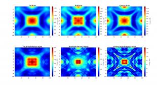
- Brain Atrophy
- Lateral Ventricle Enlargement
- Cerebrospinal fluid (CSF)
- Accumulation of plaques and tangles
- Signal Pre-Processing techniques to eliminate clutter (Skin, Skull and Antenna Coupling)
- Develop imaging algorithms to display areas of the brain affected by the disease using data acquired through simulations/sensors.
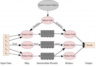
- Optimization of microwave Imaging algorithms using big data frameworks to improve efficiency.
- Investigate the use of Machine learning for the Classification of Alzheimer using microwave scattering data.
Recent Publications:
- Ullah, R., Saied, I., & Arslan, T. (2022). Measurement of whole-brain atrophy progression using microwave signal analysis. Biomedical Signal Processing and Control, 71, 103083. https://doi.org/10.1016/j.bspc.2021.103083
- Ullah, R.; Arslan, T. Parallel Delay Multiply and Sum Algorithm for Microwave Medical Imaging Using Spark Big Data Framework. Algorithms 2021, 14, 157. https://doi.org/10.3390/a14050157
- Ullah, R.; Arslan, T. PySpark-Based Optimization of Microwave Image Reconstruction Algorithm for Head Imaging Big Data on High-Performance Computing and Google Cloud Platform. Appl. Sci. 2020, 10, 3382. https://doi.org/10.3390/app10103382
- R. Ullah and T. Arslan, "Detecting Pathological Changes in the Brain Due to Alzheimer Disease Using Numerical Microwave Signal Analysis," 2020 IEEE International RF and Microwave Conference (RFM), 2020, pp. 1-4, doi.org/10.1109/RFM50841.2020.9344758


